
01
Two pumps in one fist-sized muscle
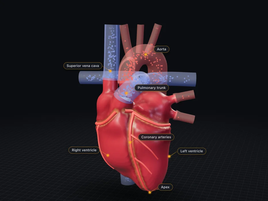
Make a fist. Your heart is roughly that size: about 12 by 8 by 6 cm. It weighs 250 to 350 g. It sits in the middle of your chest, behind the breastbone and between the lungs. It is tilted, so about two thirds of it lies left of the midline, and its tip, the apex, points down and to the left. At rest a healthy heart beats about 60 to 100 times a minute. In a study of 92,000 adults who wore fitness trackers, the average was about 65.
In a typical example, each beat pushes out about 70 mL of blood. At rest that adds up to about 5 litres a minute, roughly as much blood as your whole body holds. So on average your blood makes a full lap of the body about once a minute. Your heart beats about 100,000 times a day. The heart is really two pumps side by side. The right side sends blood through the lungs to pick up oxygen. The left side sends it on round the rest of the body.
Each pump has two rooms. An atrium collects the blood coming in, and a ventricle pushes it out. Four one-way valves keep the blood moving forward. The model shows one resting beat, at 75 beats a minute, slowed right down. It takes a few shortcuts. Oxygen-poor blood is drawn blue, as in most anatomy books, but in life it is dark red. In a real heart the atria sit more behind the ventricles than on top of them. The vessels are cut off a hand's width from the heart. Drag the timeline to move through the beat, and drag the model to turn it.
You may have heard that the heart never rests. In fact it rests in every beat. At rest the ventricles spend only about two fifths of each beat squeezing. For the other three fifths the muscle relaxes while the heart fills again. This relaxed part is called diastole, and the squeeze is called systole. There is no single normal pulse, either. In that tracker study, resting rates ran from about 40 to over 100. One healthy person's normal rate could differ from another's by up to 70 beats a minute.
02
Four rooms, two pressures
The model is now sliced open and seen from the front, a textbook view that shows all four rooms at once. It faces you, so the heart's right side is on your left. Blood that has given up its oxygen comes back from the body through two big veins, the venae cavae. They empty into the right atrium. From there the blood passes into the right ventricle, which pushes it through the lungs next door. That takes little force: its pressure peaks at about 25 mmHg.
Blood rich in oxygen comes back from the lungs through four pulmonary veins into the left atrium. From there it passes into the left ventricle, the strongest room. This one must push blood round the whole body, from your brain to your toes. Its pressure peaks at about 120 mmHg, roughly five times the right side's. So its wall is about three times as thick as the right ventricle's, typically 6 to 9 mm. You can see the difference in the cut.
A muscular wall, the septum, divides the two sides, so oxygen-rich and oxygen-poor blood do not mix. Both ventricles move the same amount, about 70 mL per beat in a typical example. If one side kept pumping even a little more, blood would pile up behind the other. Most filling happens on its own: blood flows through the atria into the relaxed ventricles. At the end, the atria squeeze and add a small top-up: up to about a fifth at rest, and more when the heart races. Tap a room to see where its blood comes from and where it goes.
How does each side know how much to pump? Nothing has to tell it. Heart muscle follows a simple rule called the Frank-Starling mechanism: the more a ventricle fills, the harder it squeezes. Extra blood stretches the muscle cells, stretched cells pull with more force, and the ventricle pushes the extra out. So what flows in flows out again, beat after beat. The rule is built into the muscle itself. Endurance athletes use it far more than untrained people: their hearts fill more, so they pump more.
03
Four one-way doors
Between each atrium and its ventricle sits a valve made of thin flaps called leaflets. On the right it is the tricuspid valve, with three leaflets. On the left it is the mitral valve, with two. When the ventricle squeezes, the rising pressure pushes the leaflets shut. Thin cords, the chordae tendineae, tie their edges to small muscles in the ventricle wall, called the papillary muscles. These tighten during the squeeze, so the leaflets cannot blow back into the atrium like an umbrella in the wind.
The exits have a different kind of valve. The pulmonary valve opens the way to the lungs and the aortic valve the way to the body. Each has three cusps shaped like small pockets. While blood rushes out, the pockets lie flat against the wall. When the ventricle relaxes, blood starts to fall back and fills the pockets. They snap shut and meet in the middle. The model shows all four valves in one cut for clarity. In a real heart the pulmonary valve sits higher and further forward.
No valve has muscle of its own. Each one opens when the pressure behind it is higher than the pressure in front, and shuts when that reverses. As the valves snap shut, they make the heart sounds, the lub-dub a doctor hears through a stethoscope. Lub, the first sound, is the mitral and tricuspid valves shutting as the squeeze begins. Dub, the second, is the aortic and pulmonary valves shutting as it ends. So the sounds come from the valves, not from the muscle. Tap a valve, and the camera follows it through the beat.
Each sound is really two. The mitral valve shuts a fraction of a second before the tricuspid, a gap too short to hear with a stethoscope. Tap each one and compare the times they shut to see the gap in the model. The aortic valve also shuts just before the pulmonary valve. That gap, called the split of the second sound, varies as you breathe and even when you change position. A healthy adult heart makes only these two sounds. A third sound is normal in children, but in adults it is often a sign of disease.
04
One beat, step by step
The graph follows the left side through one beat. It starts with the ventricle relaxed and already mostly full. The atria squeeze and top it up to about 120 mL. Then the ventricle begins to squeeze. The mitral valve shuts, and for a moment all four valves are closed. Blood has nowhere to go, so the volume stays the same while the pressure shoots up, from about 10 to about 80 mmHg. This phase has a long name, isovolumetric contraction: squeezing at the same volume.
At about 80 mmHg the ventricle's pressure passes the aorta's, and the aortic valve swings open. Blood rushes into the aorta at up to about 1 m/s, and the pressure peaks at about 120 mmHg. In a quarter of a second or so, the ventricle pushes out about 70 mL, and about 50 mL stays behind. So each beat sends out only a bit over half the blood, about 55 to 65 percent. This share is the ejection fraction. The curves are typical textbook shapes, not one person's measurement, and real hearts, when measured, often hold more.
As the squeeze fades, blood starts to fall back and the aortic valve snaps shut. That makes the little notch in the aortic curve. All four valves are shut again while the pressure falls, for 0.07 s or less. Once it drops below the atrium's, the mitral valve opens and blood flows in, fast at first, then slowly. The elastic aorta stretches with each push and springs back between them, so its pressure only falls to about 80. The high and the low together are the blood pressure in the aorta, the familiar 120 over 80.
Heart muscle has its own blood supply, the coronary arteries on its surface. But a squeezing muscle presses on its own vessels, so most of that blood flows between beats, while the muscle relaxes. When the heart speeds up, it is mostly the relaxed part that gets shorter. At 75 beats a minute the heart spends about 33 seconds of every minute relaxed. At 150 it gets only about 22. So doubling the rate cuts the heart's own feeding time by about a third. That matters most when the coronary arteries are narrowed.
05
A spark that starts inside
Every beat starts in the heart itself, not in the brain. High in the wall of the right atrium sits a small patch of special cells, the sinus node. Left to itself, it would fire about 100 times a minute. But the vagus nerve constantly holds it back, down to a resting 60 to 80. Nerves and hormones such as adrenaline only turn the rate up or down. Breathing does too. Each breath in briefly loosens the grip of the vagus, so a healthy pulse speeds up a little as you breathe in and slows as you breathe out.
Each time the node fires, an electrical wave spreads across both atria in about 90 ms, and they squeeze. On the trace this is the small bump called the P wave. Then the wave meets a ring of tough tissue that it cannot cross, except at one gate: the AV node, short for atrioventricular node. It is the slowest part of the system: the signal crawls through at about 0.05 m/s. It holds the signal for about a tenth of a second, so the atria finish emptying before the ventricles start to squeeze.
Past the node, the signal runs down the bundle of His and its two branches, one on each side of the septum, at about 2 m/s. A fine network, the Purkinje fibres, then spreads it over the inner walls at about 4 m/s, roughly 80 times faster than in the node. The whole ventricle wall fires in less than a tenth of a second: the tall spike on the trace, called QRS. Then the walls recover, and that shows on the trace as the T wave. Tap a wave to jump to that moment.
A transplanted heart shows how much the heart runs itself. It arrives with no nerves connected, yet it beats. With no vagus to slow it, it beats faster than normal at rest. During exercise it still speeds up, only more slowly, as adrenaline and similar hormones reach it through the blood. In 40 to 70 percent of patients some nerves grow back, months to years later. The heart has spare pacemakers too. If the sinus node fails, the AV node takes over at about 40 to 60 beats a minute.
06
Two loops, and what running changes
Follow one drop of blood. The right ventricle sends it through the pulmonary arteries into the lungs, where it picks up oxygen. It returns through the pulmonary veins to the left side, which sends it out through the aorta to the body. Then it comes back through the venae cavae to the right side. So there are two loops in a row, and the heart drives both at once. An artery is any vessel that carries blood away from the heart, with or without oxygen: the pulmonary arteries carry oxygen-poor blood. At rest a drop goes round in about a minute on average.
Now slide the effort up. The rate climbs from about 75 towards about 190 beats a minute for a 25-year-old, and each beat pushes out more. Heart rate times the blood per beat is the cardiac output, the blood pumped each minute. It rises from about 5 litres at rest to about 20 in hard exercise. The squeeze gets a little shorter, but the time to fill shrinks far more. When the rate doubles, the squeeze loses less than a third of its time and the filling about two thirds. Watch the blue part of the chart shrink.
All that work has a cost. The heart feeds itself through its coronary arteries, which take about 250 mL of blood a minute at rest, roughly 5 percent of what it pumps. It uses about a tenth of the oxygen your body takes up. Its pumping power is modest, though: about 1 W for the left side. Over a lifetime it beats about 3 billion times. That is a lot for a mammal. Across mammals, a lifetime comes to roughly a billion beats, within a factor of ten. A diving blue whale's heart beats only 2 to 8 times a minute.
Endurance training reshapes the heart. The left ventricle grows roomier, about 55 mm across in male athletes against about 48 mm in untrained people, and its wall thickens slightly. This athlete's heart is a healthy change. Each beat moves more blood, and the resting rate drops to about 50, nearer 30 in elite cyclists. Flat out it pumps about 28 litres a minute. The records are about 42 litres a minute and 210 mL in one beat. Yet its top rate is no higher than anyone else's. And 220 minus your age is only a rough guess, off by about 10 beats either way. Tap the athlete to compare.